ECG – How To Read An ECG
<< Back to Medical Nursing Videos
Electrocardiogram or ECG Rhythm videos (also known as EKG) can be seen below. ECG was first time developed in 1901 by a German scientist and is a measuring device that physicians use to record the normal and abnormal electrical activity. Each beat of the heart is presented by an electrical impulse and its beginning is in the upper-right side of the heart.
 The beat is transmitted through the nerve cells into the bottom right and left portion of the heart. The recurrent electrical impulse pattern is stimulating the heart muscle cells to contract and pump blood throughout the entire body.
What Is ECG or Electrocardiogram?
There are few common EKG rhythms that are represented here that you need to know about them:
(Videos on
this site are for entertainment purposes only. Please
do not attempt any of the procedures seen in these videos
without formal medical education & licensing.)
Normal
Sinus Rhythm
Normal Sinus Rhythm
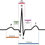
Looking at this ECG video you will probably notice that:
- The rhythm is regular
- The rate is 60-100 bpm
- The QRS Duration is normal
- P Wave is visible before each QRS complex
- P-R Interval is normal
- Shows that the electrical signal is generated by the sinus node and traveling in a normal fashion in the heart
Atrial Fibrillation
Atrial Fibrillation
Atrial Fibrillation is a common cardiac arrhythmia that causes irregular heartbeat. It is often associated with fainting, chest pain or congestive heart failure, but in some cases atrial fibrillation is caused by benign conditions.
Looking at the EKG video you can notice:
- The rhythm is irregularly irregular
- The rate is 100-160 beats per minute- if the patient is on medication the beats are slower.
- QRS Duration is normal
- The P Wave is not distinguishable
- The P-R Interval is not measurable
- The atria fire electrical impulses are in irregular fashion causing irregular heart rhythm
Ventricular Tachycardia
Ventricular Tachycardia
Ventricular Tachycardia is a rapid heartbeat and starts in the ventricles. The pulse is more than 100 beats per minute and there are at least three irregular heartbeats in a row. Ventricular tachycardia occurs in patients with: cardiomyopathy, heart failure, heart surgery, myocarditis, valvular heart disease.
Looking at this video you may notice that:
- The rhythm is regular
- The rate is 180-190 beats per min.
- The QRS Duration is prolonged.
- P Wave – you cannot see
- The poor cardiac output is related with the irregular rhythm that causes the patient to go into cardiac arrest.
Asystole
Asystole
Asystole is a state when there is no cardiac electrical activity, no contractions of the myocardium and no cardiac output or blood flow. Asystole can be treated by cardiopulmonary resuscitation (CPR) and epinephrine (adrenalin) as combination.
Looking at the video you can notice that:
- The rhythm is flat
- There are 0 beats per minute
- There is none QRS Duration
- P Wave- None
Ventricular Fibrillation
Ventricular Fibrillation
The electrical signals are disorganized and they are causing the ventricles to quiver instead of contract in a rhytmatic fashion. Ventricular fibrillation is commonly identified arrhythmia in cardiac arrest patients. These patients are treated with defibrillation.
Looking at this video you will notice that:
- There is Irregular Rhythm
- The rate is 300+ and is disorganized
- The QRS Duration is no recognizable
- P Wave- not seen
- You have to defibrillate your patient quickly!
Premature Ventricular Contraction (PVC)
Premature Ventricular Contraction (PVC)
Premature Ventricular Contraction (PVC) is also called premature ventricular complex, ventricular premature contraction, ventricular premature beat or extra systole. This is a common event where the heartbeat is initiated by Purkinje fibres in the ventricles rather than by the sinoatrial node.
Looking at this EKG video you will notice that:
- The rhythm is regular
- The rate is normal
- The QRS Duration is normal
- The P Wave rate is normal
- The P-R Interval is normal
- You will notice that 2 odd waveforms that are depolarizing prematurely in response to a signal within the ventricles.
Atrial Flutter
Atrial flutter
Atrial flutter occurs in the atria of the heart and is an abnormal heart rhythm. It is associated with tachycardia at the beginning because of the fast heart rate, and after that falls into the category of supra- ventricular tachycardias.
Looking at this video you will notice that:
- The rhythm is regular
- There are 110 beats per minute
- The QRS Duration is normal
- P Wave rate- there is 300 beats per minute
- The P-R Interval is not measurable
- The rapid heart rate is in the atria
Pacemaker Rhythm
Pacemaker Rhythm
The pacemaker rhythm can be easily recognized on the EKG video. The video shows pacemaker spikes and the vertical signals are representing the electrical activity of the pacemaker. The pacemaker rhythm shows that:
- The rhythm is regular
- There are 80 beats per minute
- The QRS Duration is normal
- P Wave is normal


